EPIQ Elite
Performance redefined


EPIQ Elite
Performance redefined
EPIQ Elite Elevate provides high-quality imaging and tailored clinical information to help clinicians deliver timely, confident answers to more patients worldwide. With advanced intelligence and an exceptional level of performance, EPIQ Elite meets the demands of today’s most challenging practices.
Clinical image gallery
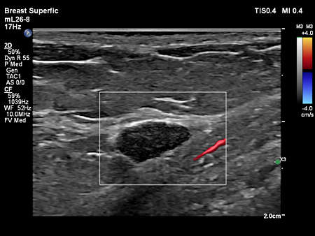
- eL18-4 scanning on a breast lesion.
- L12-3 ERGO Vascular Carotid Flow Viewer Color
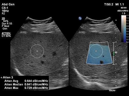
- Affiniti VM10.0 C5-1 Abd Gen Liver Fatty LFQ
- mL26-8 finger tendon
- Affiniti VM10.0 L12-5 Adv Breast 2D
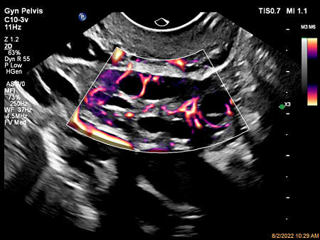
- Ovary MFI Flow Viewer
- L12-3 paired with CEUS Auto Scan, scanning on the Vascular Carotid Preset.
- mL26-8 paired with Flow Viewer, scanning on a thyroid
- Placenta CPA with Flow Viewer
- Ovarian blood flow with MFI using C10-3v transducer
- Uterus Color Flow with Flow Viewer
- mL26-8 paired with Flow Viewer, scanning on a breast lesion
- C5-1 Liver Fat Quantification
- eL18-4 paired with CEUS, scanning on a thyroid.
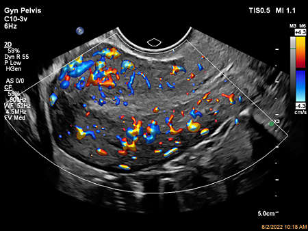
- Color Flow with Flow Viewer using C10-3v showing ovary perfusion
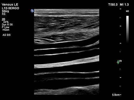
- L12-3 Ergo scanning on a calf vein
- Flow Viewer applied to MFI with C10-3v uterus
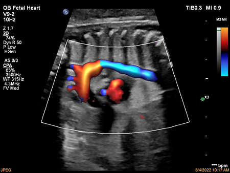
- Fetal Heart CPA with Flow Viewer
- Fetal Aortic Arch CPA with Flow Viewer
- Linear ElastQ Breast
- C5-1 paired with Flow Viewer, scanning on the Pediatric Abdomen
- Umbilical cord shown with Color Flow with Flow Viewer using C5-1 transducer
- Strain elastography demonstrating increased stiffness of an intrauterine fibroadenoma
- Pulmonary vein and fetal heart MFI with Flow Viewer using C5-1 transducer
- Affiniti VM10.0 L12-5 Adv Breast 2D
- Aortic arch CPA with Flow Viewer using C9-2 transducer
- eL18-4 Vertebral Imaging
- Fetal Lung Perfusion Color Flow with Flow Viewer
- Ovary Color Flow with Flow Viewer
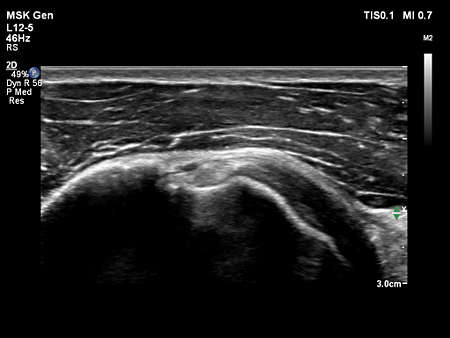
- Affiniti VM10.0 L12-5 MSK Gen Biceps Tendon
- C9-2 scanning on the Abdomen Renal Preset
- mL26-8 Pediatric Liver
- Auto Scan Of
- Umbilical Cord Color Flow with Flow Viewer
- C9-2 scanning on a liver.
- L12-3 Ergo paired with Flow Viewer, scanning on a carotid
- MSK Superfic Bowel FV
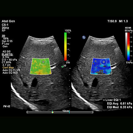
- C5-1 Liver ElastQ
- Fetal heart four-chamber color flow with Flow Viewer


Features
Auto ElastQ
Perform automated liver elastography with Auto ElastQ, and experience our next generation of liver health assessment. Auto ElastQ is designed to simplify user workflow with real-time, quantitative shear wave measurements.
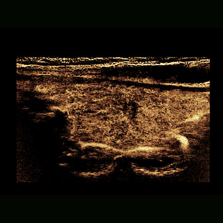
CEUS high frame rate linear & Auto Scan
See a 67%*** increase in CEUS frame rate and a 76%*** increase field of view with the eL18-4 transducer when thyroid scanning. CEUS Auto Scan improves image uniformity and sensitivity

Contrast-enhanced ultrasound (CEUS)
CEUS can transform the role of ultrasound in the liver, allowing the study of the enhancement patterns of suspicious liver lesions in real time, as well as provide an alternative non-ionizing approach to the assessment of vesicoureteral reflux in pediatric patients.
Specifications
- Common Specifications
- Width
- 60.6 cm/ 23.9 in
- Height
- 146-171.5 cm/ 57.5-67.5 in
- Depth
- 109.2 cm/ 43 in
- Weight
- 104.3 kg/ 230 lb without peripheral devices
- Control panel
- Monitor size
- 24 inch / 60.96 cm HD display
Related products
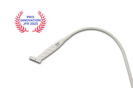
- Discover the award-winning Philips mL26-8 ultra-high frequency compact linear array transducer, designed to provide exceptional imaging versatility from head to hip. With specialized presets for MSK, breast, vascular, dermal, and ocular applications, the mL26-8 offers unmatched adaptability on EPIQ & Affiniti. Proud recipient of the 'Best Innovation Award in General Imaging' at Journées Francophones de Radiologie 2023.
Disclaimer
Available in select countries. Please consult your Philips representative for further details.
*based on a sample size of 20 users
***Compared to previous capability