- Featuring
- July 11 2024
- 10 min read

Using MRI to help identify suspicious lesions in patients with suspected prostate cancer
WellSpan York Hospital, based in York, Pennsylvania, USA, is an advanced specialty care hospital. According to Dr. Steiner, the Chairman of Imaging, the hospital has been using multiparametric MRI and fused MRI/ultrasound image-guided biopsies in prostate cancer diagnosis since mid-2019. He has built a referral base within the five-county community surrounding York and northern Maryland regions.
“Contrary to other techniques, MRI allows for examination of the entire prostate gland with high soft-tissue contrast. Multiparametric MRI allows us to identify suspicious lesions and give these a PI-RADS score [1]. For high-risk lesions, a biopsy can then be performed, guided by MRI images that are fused with ultrasound images in real time. This allows targeting of the lesions that were identified before.”
The power of multiparametric MRI is now not only recognized for exams to identify suspicious lesions, but also expands into guiding biopsies to inform a definite diagnosis.

Edward Steiner
Chief and Medical Director
WellSpan Advanced Prostate Care Center, York, Pennsylvania, USA
Advanced multiparametric MRI helps clinicians boost prostate cancer diagnosis
Dr. Steiner explains that techniques for prostate imaging and cancer diagnosis have not changed substantially in the past 30 years, despite the known limitations. PSA testing alone is usually insufficient and current TRUS biopsy techniques often miss anywhere from 40 to 50% of the gland [2]. “PSA testing is imprecise and has a significant number of false positive as well as false negative tests,” he says. “It is, however, the accepted first path of entry for most patients that are ultimately diagnosed with prostate carcinoma.”
The use of MRI has significantly improved capabilities in prostate cancer diagnosis, according to Dr. Steiner. “Multiparametric prostate MRI allows us to look at three parameters to build our diagnosis on: conventional T1 and T2 signal intensity, diffusion-weighted imaging and ADC map, as well as dynamic flow imaging, to define the highest probability of prostate carcinoma.”
The standard PI-RADS system is then used to grade lesions based on the MRI findings. For PI-RADS 1 and 2, clinically significant cancer is (highly) unlikely. Intermediate PI-RADS 3 lesions represent a kind of diagnostic “grey area” – these lesions may become PI-RADS 4 lesions if they demonstrate a fusion restriction and a hypervascular tumor flow pattern or depending upon index of suspicion. PI-RADS 4 and 5 lesions have a statistically high chance of being a clinically significant prostate carcinoma and should be biopsied. Once the biopsy is performed, the pathologists characterize the biopsy samples with either a Gleason score or an ISUP grade group [3].
“For lesions in the prostate’s peripheral zone, the DWI (diffusion weighted imaging) and ADC map are most helpful for our diagnoses, says Dr. Steiner. “ In this case, the DWI shows very bright signal, which indicates diffusion restriction. The arcuate area with significant signal drop out (arrow) on the ADC map is recognized as highly suspect for tumor. On the axial T2- weighted image the capsule contour looks a little irregular (arrow), which we interpret as capsular disruption and I usually give a measurement: this lesion shows larger than 1.5 cm capsular disruption. I don’t see any signs of lymphadenopathy but interpret this lesion as PI-RADS 5. The hyper vascular flow pattern in the bottom images adds to the diagnostic confidence”.
Predictable MRI patterns help identify suspicious lesions in the whole gland
Depending upon the Gleason score and prior therapies, prostate carcinoma has a certain predictable pattern on multiparametric MRI, according to Dr. Steiner. “In general, lesions in the peripheral zone have decreased T2-weighted signal and are relatively focal,” he says. “In the transitional zone, these lesions are more difficult to evaluate on T1 and T2, but are generally non-encapsulated.”
“We especially look at diffusion-weighted images and the ADC map. Prostate neoplasms generally have diffusion restriction, so they are bright on diffusion-weighted imaging and dark on an ADC map, which is one of the most important characteristics of neoplasms.”
“The third characteristic we look at, flow, is somewhat less specific but may be quite important in deciding whether a lesion is significant or insignificant. Prostate neoplasms often have a hypervascular tumor flow pattern, meaning that there is rapid inflow of blood into the lesion and then rapid outflow due to a disrupted capillary bed. This can be graphed on multi-parametric images, allowing us to define regions of interest and look at the actual flow within these regions.”
“I perform this interrogation using DynaCAD prostate, which also provides an easy way to determine PI-RADS score and create the report for the urologist.”
Using detailed, segmented MRI images to guide prostate biopsy
Dr. Steiner explains how a “blind” ultrasound biopsy may lead to a negative result, even when a tumor is present. “In a non-targeted biopsy guided by ultrasound, you see the needle and the confines of the prostate, but cannot see the tumor. So, when trying to get 12 cores as evenly distributed as possible, the tumor may still be missed, particularly when it is in the anterior gland, low in the apex or in other regions generally not easily biopsied by ultrasound.”
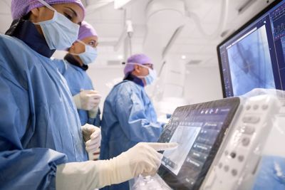
That is why Dr. Steiner has implemented the pathway where the MRI images can also be used to guide the biopsy. He uses an MR/ ultrasound fusion guided biopsy system, UroNav, which fuses pre-biopsy MRI images of the prostate with real-time ultrasound images during transrectal biopsy, for excellent delineation of the prostate and suspicious lesions, as well as clear visualization of the biopsy needle path.
“I felt strongly that the urologists are used to doing free-hand biopsies – their brain and hand are very used to manipulating the probe,” says Dr. Steiner. “What UroNav offers is no change in that workflow; it takes the diagnostic MRI images and the localized, segmented lesions and adds tracking and navigation to fuse that with the live ultrasound images. In this way, the MRI images can be used for targeting the lesion when performing the biopsy. The UroNav navigation sensor is mounted on the TRUS probe*, so for urologists the manipulation is similar to what they were used to.”
“This process allows us to perform focal biopsies of suspicious areas based on PI-RADS categories that indicate the probability of an underlying potential malignancy,” says Dr. Steiner. “As a result of the high confidence gained using this pathway, I have in the meanwhile limited my biopsies to fewer than ten and my goal, as I’m getting more comfortable with the process, is six or less,” he says.
"We generate over 4,500 images per case. Not utilizing an automated process would make interpretation quite difficult."
Copy this URLto share this story with your professional network
Disclaimer
Results are specific to the institution where they were obtained and may not reflect the results achievable at other institutions. Results in other cases may vary.